A New Standard for Liver Disease Detection
“A New Standard for Liver Disease Detection” by MD News
In the last five years, magnetic resonance elastography (MRE) has emerged as a highly effective, noninvasive method of measuring hepatic fibrosis in the setting of diffuse liver diseases such as hepatitis B, hepatitis C and non-alcoholic hepatosteatosis (NASH). St. Paul Radiology specialists collaborate with the developers of MRE at the Mayo Clinic–Rochester, bringing this cutting–edge technology to the Twin Cities area.
According to Peter Wold, M.D., the stiffness, or elasticity, of the liver acts as a “surrogate marker for hepatic fibrosis.” If inflammation caused by diffuse liver disease persists, hepatic fibrosis may develop. Hepatic fibrosis may progress to cirrhosis, stage IV fibrosis, which places the patient at risk for development of liver failure, hepatocellular carcinoma and complications of portal hypertension (e.g., varices). Traditionally, Dr. Wold says, fibrosis has been staged using a numerical scale (0, I, II, III, IV) of increasing severity. Stage 0 represents no fibrosis and stage IV is end–stage fibrosis (i.e., cirrhosis). Biopsy remains the “gold standard” for staging liver fibrosis, but has significant drawbacks, such as high cost, potential for bleeding, small sample size and need for sedation. MRE is a reproducible, noninvasive test to measure liver elasticity and provides accurate imaging data to stage the severity of diffuse liver disease.
“The purpose of the MRE is to see if the liver is abnormally stiff,” Dr. Wold says. “If so, how much? It’s very much a niche examination. It’s designed for people who have some type of diffuse liver disease — such as hepatitis B, hepatitis C or hepatic steatosis — and not for someone who has a mass, such as a tumor, in his or her liver.”
MRE: A Procedural Overview
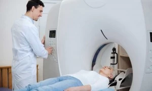
MRE techniques can be performed on a magnetic resonance imaging (MRI) machine, modified with an acoustic driver and special software, according to a study published by Talwalkar Et Al. in the journal Hepatology. The study explains that the acoustic driver sits outside the magnet room — the chamber where the patient lies during the MR scans — and generates “acoustic pressure waves” between 40 and 120Hz per second. A tube connecting the wave–generating driver to another driver placed on the patient’s anterior abdominal wall transfers the waves.
“The waves cause tiny cyclic displacements as they propagate,” says Richard L. Ehman, M.D., author of Magnetic Resonance Elastography: An Emerging Tool for Cellular Mechanobiology, and MRE pioneer. “Cyclic displacement” is measured and imaged, Dr. Wold explains, by specialized software that converts data into kilopascals. Results help determine if a patient’s fibrosis has progressed to the point where antiviral treatment should be administered, or if cirrhosis has developed.
Why Biopsy Just Doesn’t Cut It
Nezam H. Afdahl, M.D., says that traditional biopsy methods can be accurate in diagnosing healthy and cirrhotic livers, but the stages in between can be problematic due to biopsy size, sampling error and varying opinions of the analysis in “Staging Liver Fibrosis: Liver Biospy, Imaging and Biomarkers,” published by the American Gastroenterological Association (AGA). Rouviere et al.’s study “MR Elastography of the Liver: Preliminary Results,” published in the journal Radiology concluded MRE to be an accurate method of ascertaining liver stiffness and diagnosing all stages of fibrosis.
Although biopsies can be effective and safe, the procedure’s repeatability is a significant drawback. Patients who are at risk for fibrosis or cirrhosis may need frequent monitoring, so the development of an alternative diagnostic scan to biopsy that avoids the additional risk of bleeding and requires sedation, all while sampling the entire liver — not just a 2 cm core tissue sample — is a coup for both patients and physicians.
St. Paul Radiology is on the cutting edge of this new technology. The practice’s Body–Imaging Section, a unit of radiologists with fellowship training in gastrointestinal (GI) training, has conducted 528 MRE exams, according to Armen Kocharian, Ph.D., St. Paul Radiology’s medical physicist. As the word has spread about MRE, Dr. Wold says, the number of biopsies St. Paul Radiology performs has dropped because of the appealing noninvasive nature of MRE.


