


Arteriovenous Malformation (AVM)
ProceduresWhat is an AVM?
An arteriovenous malformation (AVM) is a congenital abnormality of blood vessel development resulting in a direct communication between arteries and veins. The normal arrangement is a capillary bed intervening between an artery and a vein, allowing for tissue perfusion and a dissipation of pressure by the time the blood reaches the vein. In an AVM, both features are lost and an abnormal tangle of blood vessels called a nidus forms. This results in a high-flow direct shunting of arterial blood into the venous system. The immediately adjacent brain tissue may be chronically damaged from a lack of normal perfusion. The veins are typically abnormally dilated because they are exposed to pressure that they are not designed for handling.
What are the symptoms of an AVM?
AVMs may present in a variety of ways. Because of the high flow shunt, aneurysms may form on the artery supplying the nidus, within the nidus, or on the veins. If one of these aneurysms ruptures, bleeding may occur in the brain itself (intraparenchymal hemorrhage) or into the fluid space surrounding the brain (subarachnoid hemorrhage). This would typically be accompanied by a severe headache and/or neurologic deficit depending on the location of the injury. Alternatively, because of tissue that may be chronically damaged associated with the nidus/shunt, this may trigger seizures.
How is an AVM diagnosed?
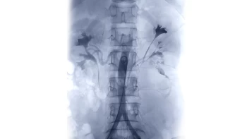
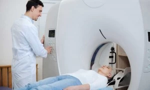
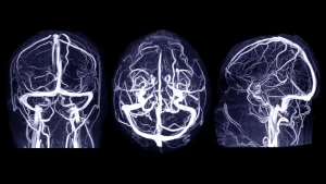
AVMs are generally readily diagnosed on standard neuroimaging (CT/CTA, MRI/MRA). Occasionally, the blood clot may compress the AVM or the AVM may be quite small and a diagnostic cerebral angiogram may be necessary for higher resolution visualization. If a lesion is not detected, repeat imaging is often performed at a delayed time point once bleeding and associated pressure has resolved.
How is an AVM treated?
Treatment of AVMs may be very complex depending on the presenting features, the anatomic area involved, and the structure of the AVM. A bleed associated with an AVM is well-established as requiring proactive intervention to prevent re-bleeding from occuring, which may be catastrophic.
A diagnostic cerebral angiogram is performed to evaluate for an aneurysm associated with the AVM. This is considered an ‘unstable’ feature that must be treated urgently. Otherwise, a complex multidisciplinary discussion is necessary. Patients achieve the best outcomes with a neurosurgeon who has dedicated experience in open surgery for AVMs, a neurointerventional surgeon highly experienced in endovascular embolization, as well as experts in neurocritical care, neurology, and potentially radiosurgery.
A treatment plan may involve surgery, radiosurgery, and/or endovascular embolization in combination.