


Calcifications
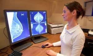
Radiologists look for tiny white specks within the breast tissue on a mammogram. These are called calcifications. They are extremely common and usually benign (non-cancerous). Widely scattered or larger white spots are normal within the breast.
Some causes for calcifications include, but are not limited to:
- Previous surgery
- Trauma
- Inflammation
- Radiation therapy
- Fibrocystic changes and other benign processes
There is no known link between calcium intake/supplements and the formation of breast calcifications.
Calcifications that are in groups or clusters require further magnified mammographic views. The radiologist will then look at their features to categorize them. These categories are as follows:
Stable calcifications
Calcifications that have remained unchanged in appearance over long periods of time in comparison to previous mammograms.
Benign calcifications
These are described as layered, tea-cup shaped, or milk of calcium. These are from benign causes such as fibrocystic breast changes or ruptured breast cysts. Some calcifications are caused from an old injury or infection in the breast tissue; fat necrosis is the term used for these. Calcifications within the skin are not concerning for a breast cancer, nor are well rounded, uniformly shaped calcifications.
Suspicious calcifications
Calcifications that have irregular shapes; these may be very faint or tiny, and may form a branching pattern. These can be more concerning for an underlying cancer, and only a biopsy of this area can determine its cause.
Indeterminate calcifications
Any calcifications that do not clearly fit into any of the categories above. The radiologist will decide if a short-term follow-up mammogram is indicated (within four to six months) to ensure calcifications are stable. However, often a biopsy will be indicated to establish the exact cause for the calcifications.
Of the calcifications that are biopsied:
- 80-90% are benign
- 10-20% prove to be a cancer
Most of these early cancers can be successfully treated by surgery and radiation with an overall survival rate exceeding 95% at five years.
Biopsy Options
The choices for a biopsy of the calcifications are either surgical (wire localization) or a less-invasive needle core biopsy (stereotactic biopsy). Both procedures have equal accuracy for diagnosis but require different preparation. The Breast Center of Suburban Imaging has brochures on these options. The Breast Center staff will keep your doctor informed of the results of your breast imaging exam(s) and of your biopsy findings. The pathology results are usually available within 24 to 48 hours. Occasionally, special tests are required and may delay the final report. The Breast Center nurse or your doctor can keep you informed if any delays should occur.