


Diagnostic Cerebral Angiography
What is a diagnostic cerebral angiogram?
A diagnostic cerebral angiogram is the gold standard for evaluation of the blood vessels of the brain. While CT and MRI scans have improved considerably over time, they have limited spatial and temporal resolution to fully define a range of neurovascular conditions. A cerebral angiogram provides exquisite detail that is often required to make a diagnosis and define the intricate anatomy that dictates the best treatment.
How is a diagnostic cerebral angiogram performed?
Generally, a diagnostic cerebral angiogram is an elective, outpatient procedure, although it can also be performed on an inpatient basis. Patients are sedated for the procedure in addition to using local anesthesia.
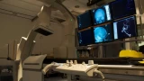
A small needle is then used to access the artery in the right groin area (femoral artery). Occasionally, the radial artery in the wrist is used for access. A catheter is then navigated through the arterial tree of the body to the blood vessels of the neck. An iodine-based dye is then injected through the catheter and high-resolution x-ray images of the brain blood vessels are then acquired. For certain conditions, a 3D image is also obtained.
After the procedure, the catheter is removed and patients recover for approximately 4 hours in the hospital before returning home.