


Dural Arteriovenous Fistula (Dural AVF) Embolization
What is a dural AVF?
Dural AVF is caused when the arteries in the brain are connected to veins not by normal capillaries but by arteriovenous fistulas. These fistulas can empty the arteries of blood before cerebral circulation is completed, causing headaches, hemorrhage, seizures, and strokes. They can also cause neurological symptoms impacting memory, movement, vision, or speech.
Endovascular embolization is a minimally invasive technique performed to cut off the blood supply to a specific part of an artery or vein, and is an effective treatment for the vast majority of patients.
During endovascular embolization, a catheter is passed through the groin up into the arteries or veins in the head that lead to the DAVF to inject a glue-like material or coils. This injection shuts off the flow of blood through the DAVF.
We evaluate each DAVF patient to decide the best treatment for that patient's specific DAVF. In special cases, we may opt to use alternative treatments, or a combination of techniques.
What are the symptoms of a dural AVF?
While these lesions may be entirely asymptomatic, the most characteristic presentation is pulsatile tinnitus - a rhythmic whooshing sound from the turbulent blood flow of the fistula that propagates through the bone of the skull. A wide variety of additional symptoms may be seen depending on the location of the fistula and the brain structures affected.
How is a dural AVF diagnosed?
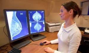
The diagnosis of a dural AVF is often suggested on a CT or MRI scan of the blood vessels of the brain. This is then further confirmed with a diagnostic cerebral angiogram, which demonstrates the dynamic blood flood of the fistula and its fine anatomic detail.
How is a dural AVF treated?
The current gold standard for treatment of dural AVFs is endovascular embolization. There are a number of different endovascular devices, techniques and materials that can be applied to best treat a given lesion depending on its specific configuration. A dural AVF may be treated through the artery, through the vein, or even both. Once access to the fistula is obtained, it is closed with coils and/or a glue-like substance called Onyx, eliminating the abnormal communication.