


Carotid Angioplasty/Stenting
What is carotid stenosis?
Carotid stenosis refers to a buildup of plaque in the carotid artery in the neck. This is associated with a number of risk factors including hypertension, diabetes, hyperlipidemia and smoking. Plaques may remain small and asymptomatic. Others may be very extensive and lead to significant narrowing of the carotid artery, restricting blood flow. Furthermore, blood clots may form on the plaque and subsequently travel into the arteries of the brain and cause a stroke. Even plaques with relatively mild degrees of narrowing can have unstable features and break down, causing clots to form.
What are the symptoms of carotid stenosis?
Symptoms of carotid stenosis range from transient ischemic attacks, in which the patient experiences a temporary neurologic deficit, to an extensive, debilitating or ultimately fatal stroke. One well-described temporary neurologic deficit is known as amaurosis fugax. This occurs due to temporary interruption of blood flow to the eye from small clots, in which patients experience transient blindness as if a shade were being pulled down over their eye.
How is carotid stenosis diagnosed?
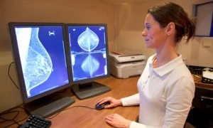
Carotid stenosis is readily diagnosed by a variety of imaging modalities including ultrasound, CT and MRI. These studies allow us to determine the presence of plaque, the severity of narrowing it causes, and in some cases the composition of the plaque/particular features that may elevate the risk of stroke.
How is carotid stenosis treated?
There are several options for treating carotid stenosis, all of which have variable criteria. We will focus here on the endovascular procedure that we perform called carotid stenting. Open vascular surgeons perform carotid endarterectomy or TCAR (transcarotid revascularization), the latter often in collaboration with a neurointerventionalist experienced in carotid stenting and catheter-based acute stroke treatment. Carotid stenting and carotid endarterectomy have similar safety and effectiveness profiles, but applications and practice patterns vary. Patients that have had prior neck surgery or radiation therapy often are referred to carotid stenting given challenges with tissue instrumentation or healing after surgery in this context. Conversely, patients with difficult anatomy for endovascular catheter navigation or stent placement are best served with open surgery. Carotid stenting is performed under local anesthesia. Patients are at most mildly sedated in order to monitor for any neurologic deficit during the procedure. The arterial system is accessed either via the femoral artery in the groin or the radial artery in the wrist. The stenosis is carefully evaluated with detailed imaging. Rarely, the stenosis appears worse than it actually is on the preceding CT or MRI study, and may not be a candidate for stent placement if it is only mild to moderate in degree on high-resolution angiographic images during the procedure. Once severe stenosis is confirmed, the stenosis is crossed with a small device that opens into a cage beyond the stenosis. This is a distal embolic protection device to capture any clot that arises from the plaque during manipulation. The stent is then placed to open up the stenosis. Typically, a balloon is inflated to help open up the stent after it is deployed. Occasionally, the balloon is used prior to stent deployment if the stenosis is too tight to pass the stent catheter. Any carotid manipulation may irritate a small receptor in the region that helps to control blood pressure. Medications are administered to blunt this effect, but patients may have a sustained drop in blood pressure requiring monitoring in the ICU overnight. Rarely, this may cause a heart arrhythmia requiring immediate treatment. The catheter system is removed and a follow-up ultrasound of the neck is performed the following day and at 6 months after discharge. Further follow-up intervals are determined by the appearance at 6 months. Stenosis may recur in a minority of patients. In this instance, balloon angioplasty is performed in the same manner described to reopen the stent.